Light (LM), Transmission Electron (TEM) and Scanning Electron Microscopes (SEM)
Resolution and Magnification
Staining
Investigating cells in blood smears
Using a haemocytometer
Eukaryotic and prokaryotic cell ultrastructure
Cell plasma membranes
Light (LM), Transmission Electron (TEM) and Scanning Electron Microscopes (SEM)
One of the unifying concept in biology, that is the cell, has been arrived upon in big part thanks to microscopy. Additionally, microscopes have enabled the accumulation of knowledge about the cellular components i.e. organelles. Microscopes use visible light, as well as electrons and lasers to produce images of very small specimens prepared on slides, often dissected in a specific manner and stained with dyes, some fluorescent.
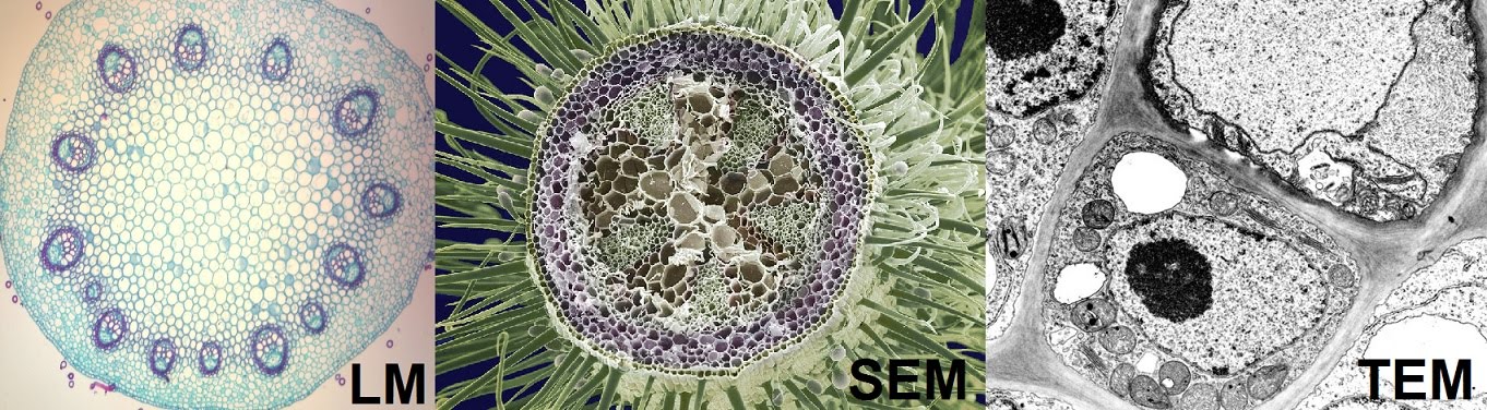
You will need to know about the difference between light, transmission electron and scanning electron microscopes – LM, TEM and SEM. Both the latter (as the name suggests) use a beam of electrons, rather than light, to produce an image of the sample.
TEM uses electrons which pass through the sample, so the resulting micrograph (image) shows everything within the sample in black and white, for example organelles in a cell. SEM uses electrons which scan the sample in 3D, resulting in a coloured micrograph with 3D detail, but no components from within the sample.
In light microscopy, light does go through the sample, but the outcome depends on the thickness of the sample. For example, the plant root slice in the diagram (LM) is thin enough to be able to see through the thickness of the sample. Light would also travel freely through air but not various materials of high opacity.
Resolution and Magnification
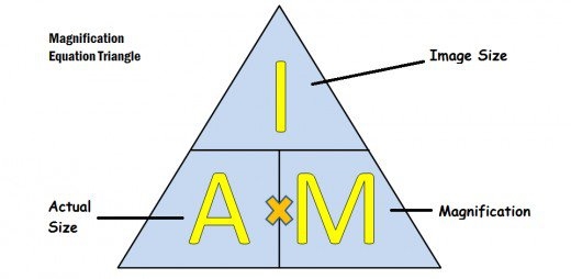
When talking about microscopes, differentiating between resolution and magnification is important. In principle, it’s not hard to understand. Imagine zooming in a photo to try to see a detail. That is magnification. Now imagine the photo has a low resolution, and if you magnify it, you can only see annoying pixels. If the image had a high resolution, you would be able to see the detail clearly after zooming in. So magnifying is zooming in, while resolution is the focus power. You will need to be able to calculate actual sizes and magnifications of various drawings. The equation for that is Image size on paper = Magnification x Actual size. This gives magnification = image size on paper / actual size. “I AM” summarises it nicely in a triangle.
Staining
Staining is a key precursor to microscopy. Most samples would not register well under a microscope without some form of staining. This can also be critical to the experiment carried out. For example, we might need a stain for the cell nucleus as well as a stain for the cell fibres…