Introduction
Immunoassays
Fluorescent labelling
Making monoclonal antibodies
Introduction
The high specificity of the antigen-antibody bond makes use of antibodies as a lab technique high on the list. Antibodies are used with proteins as a labelling method to detect presence of the target moiety (in biochemical reactions, testing or disease detection), or with whole tissue samples to detect presence of specific organelles for visualisation under a microscope.
Immunoassays
Assays are carried out in multi-well plates to detect the presence of a specific antigen, against which an antibody is added. If the antibody binds to the antigen, it causes a shift in a reporter enzyme added or linked to it, resulting in a colour change quantifiable by a spectrophotometer.
Antigens are any identifiable, specific parts of a molecule, and can be found on proteins, tissue samples, patient samples, pathogens, etc. Antibodies recognise antigens specific to them. The antigen-antibody relationship is at the heart of an organism’s self-identification and identification of invading species.
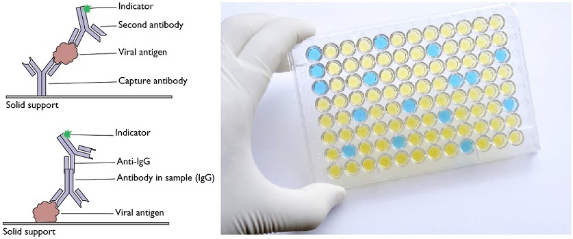
The enzyme-linked immunosorbent assay (ELISA) is a common immunoassay used to detect antigens in samples. It relies on adding the samples to a multi-well plate made of polystyrene, that can immobilise the antigen-containing molecules. The specific antibody is then added and allowed to bind, if it is to bind. After washing away the unbound molecules, the reporter enzyme is added to produce a colour change. Voila!
The antibody for the antigen is the primary antibody, while the “second antibody” binding the primary antibody (if applicable) is the secondary antibody. The latter can be the one with the indicator attached that produces a colour change.
Fluorescent labelling
The principle of primary antibody and secondary antibody is key to fluorescent labelling. The primary antibody selects the target…