Introduction
Various components of the heart
Control of heart rate
Interpreting heart function data
Investigating heart health using angiography
Introduction
In my quest to find a suitable diagram for the heart, this is what I found:
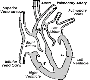
Definitely use your textbook as a guide on this. It only takes a google search to realise the ridiculous number of variations of diagrams for the heart and different annotations.
Various components of the heart
The heart contains two atria, two ventricles, the septa, AV-valves (tricuspid and bicuspid), chordae tendinae and papillary muscles. Septa describe the dividing walls between the right and left atria and ventricles respectively.
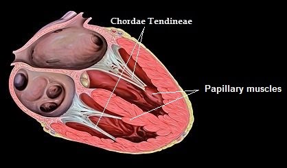
The chordae tendinae a.k.a. heart strings, are attached to the papillary muscles which prevent the walls collapsing onto themselves during heart contraction.
You need to be able to sketch a heart and label the main veins, valves, arteries and aorta, and the ventricles and atria.
There are two types of circulation going on via the heart: pulmonary circulation and systemic circulation. Pulmonary circulation is a short-distance route between the heart and the lungs, where deoxygenated blood is taken to be replenished with oxygen. Although normally veins take blood away, and arteries take blood to, in the case of pulmonary circulation things are the opposite way around. The pulmonary vein brings freshly oxygenated blood into the heart – left atrium -, while the pulmonary artery takes….