Introduction
Microscopic matrix
What is the process?
Introduction
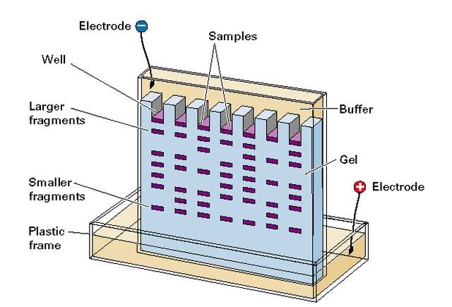
A common method of visualising differences is gel electrophoresis which involves loading small volumes of samples on a gel and running a current across it in order to separate the samples by size.
Microscopic matrix
Since the gel has a microscopic matrix inside that provides resistance against sample movement through it, the larger molecules move more slowly while the smaller fragments can move more quickly.
What is the process?
The positive charge is at the bottom of the tank, while the samples are loaded at the top. This way, they will move downwards towards the bottom of the gel because they have a negative charge as molecules. The current is run across the gel for around 30-60 minutes (ensuring the samples don’t run too long and hence run off the gel into the buffer solution! if that happens they are lost) after which…