Introduction
Transmission electron microscope
Scanning electron microscope
Light microscopy
Resolution and magnification
Staining
Introduction
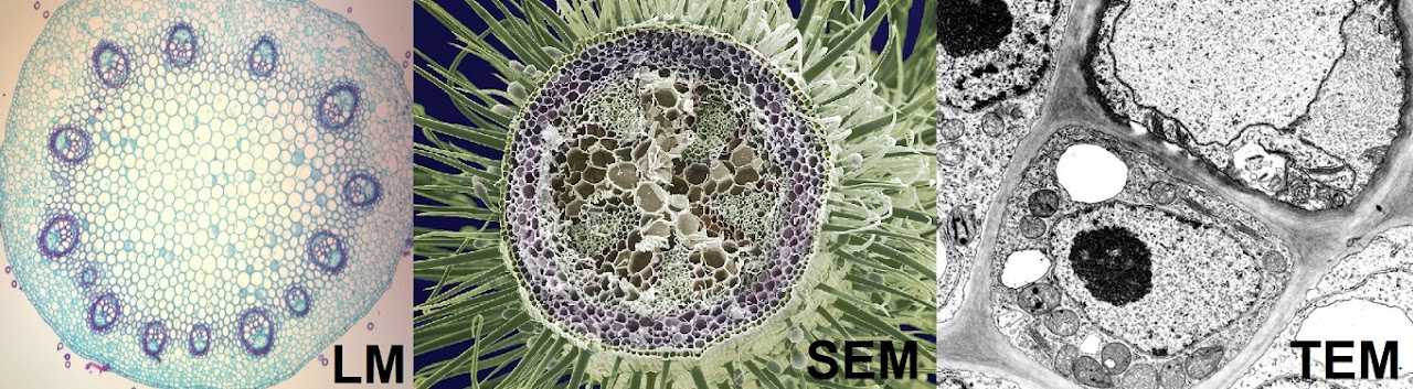
You will need to know about the difference between light, transmission electron and scanning electron microscopes – LM, TEM and SEM. Both the latter (as the name suggests) use a beam of electrons, rather than light, to produce an image of the sample.
Transmission electron microscope
TEM uses electrons which pass through the sample, so the resulting micrograph (image) shows everything within the sample in black and white, for example organelles in a cell.
Scanning electron microscope
SEM uses electrons which scan the sample in 3D, resulting in a coloured micrograph with 3D detail, but no components from within the sample.
Light microscopy
In light microscopy, light does go through the sample, but the outcome depends on the thickness of the sample. For example, the plant root slice in the diagram (LM) is thin enough to be able to see through the thickness of the sample. Light would also travel freely through air but not various materials of high opacity.
Resolution and magnification
When talking about microscopes, differentiating between resolution and magnification is important. In principle, it’s not hard to understand. Imagine zooming in a photo to try to see a detail. That is magnification. Now imagine the photo has a low resolution, and if you magnify it, you can only see annoying pixels. If the image had a high resolution, you would be able to see the detail clearly after zooming in. So magnifying is zooming in, while resolution is the focus…