Mammalian vision
Introduction
What makes up an eyeball?
The retina and photoreception
Introduction
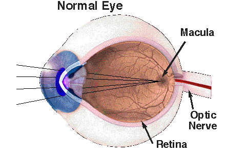
The eyeball is a magnificent contraption of brain tissue that protrudes during development to form said ball. It works to capture and process photons in light, and transmit the compressed information via the optic nerve to the visual cortex in the brain. This journey starts in the retina, which is the innermost layer at the back of the eye containing the light receptors and cells involved in this process.

Having two eyes provides binocular vision which enhances depth perception, gives a wider visual field and better distance perception.
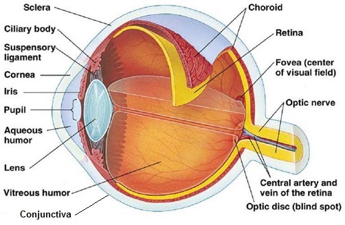
The eye has many different components that work in unison to enable vision. On its surface and lining the eyelids, the conjunctiva secretes mucus and tears, lubricating the eye and providing immune surveillance via the many microvessels.
What makes up an eyeball?
The sclera is the white of the eye and contains collagen and elastic fibre that protects the eye.
The ciliary body allows the eye to focus, while the suspensory ligament ensures the eye doesn’t fall downwards. The ability to focus is given by the added curvature given to the lens. The cornea covers the central part of the eye and refracts light, giving the eye’s optical power. Alongside the cornea, the lens, of course, refracts light.

The pupil allows the light to strike to retina, with the amount controlled by the contracting iris.
The aqueous humour is replaced frequently and consists of a plasma-like solution without proteins. On the other hand, the vitreous humour is a gel and does not get refreshed. As such, debris can accumulate in it, causing floaters. With age, it can also liquefy and collapse.
The choroid supplies the retina with oxygen and nutrients via the blood supply. Additionally, it contains pigmented cells (with melanin) to prevent internal reflection. The retina contains photoreceptor cells like cone cells which perceive visual information. This is transmitted to the brain via the optic nerve. The optic nerve creates a region without photoreceptor cells, called the blind spot because one cannot perceive images through it. The fovea is an area dense in cone cells, and serves as the key processor of central vision. This vision provides high focus on detail e.g. in reading….


